
Last updated: 2026-10-22
Hair Isn't Falling Out, It's Getting Thinner?
Androgenetic alopecia is a condition in which hair follicles gradually shrink, so the hair that grows back becomes thinner and shorter. For example, a male office worker in his 40s might compare photos from a few years ago with how he looks now and notice that, even though the number of hairs he sheds each day hasn't changed much, the hair at his crown and on both sides of his forehead has thinned into fine, short, vellus-like hair, leaving the scalp clearly visible underneath. As the condition progresses, follicle miniaturization, the shrinking of the hair follicles, continues, and the actual number of follicles still producing hair also declines (Liu Yingzi et al., 2025).
In male pattern hair loss, androgen signaling acts continuously on genetically sensitive hair follicles, which is why this miniaturization process keeps advancing. When the enzyme 5-alpha reductase in the body converts testosterone, a male hormone, into dihydrotestosterone (DHT), this DHT in the scalp binds to the hair follicles and directly causes the follicle miniaturization seen in male pattern hair loss (Kaufman K D et al., 1998).
As follicles shrink, the hair growth cycle changes along with them. Normally, the growth (anagen) phase, in which hair grows a little over 1 cm per month, makes up about 85-90% of all hair. This is followed by a catagen (regression) phase of about 1-2 months, during which growth stops (roughly 1% of hair), and then a telogen (resting) phase lasting three to four months up to a year, during which the hair stays in place before shedding (roughly 10-15% of hair), before the cycle moves back into a new growth phase. But when the follicles shrink, this cycle speeds up and the growth phase, when hair grows thick and long, shortens. As a result, newly grown hair cannot fully mature and comes in thin and short, while the catagen and telogen phases grow relatively longer, so the hair falls out more easily. Beyond this, as the number of cells in the dermal papilla, the root portion of the follicle, decreases, follicle miniaturization occurs; this can unfold gradually over many years, but it can also progress sharply within a single hair cycle (Whiting D A, 2001).
This sensitivity of the hair follicles is closely tied to genetic background. Large-scale genomic studies have identified more than 380 gene loci linked to androgenetic alopecia, including the androgen receptor pathway and the WNT signaling pathway involved in hair follicle regeneration. An epidemiological study of Korean patients similarly found a family history of hair loss in 48.5% of male patients and 45.2% of female patients (Paik J H et al., 2001). Female pattern hair loss, however, commonly occurs even in women whose blood androgen levels are completely normal. So its cause cannot be considered entirely the same as in the male pattern, and a more complex set of pathways is involved (PMID 30785992).
Men Recede from the Hairline, Women Widen from the Part
In male pattern hair loss, the hairline on both sides of the forehead typically recedes into an M-shaped pattern, and hair thins at the crown. The Hamilton-Norwood classification, widely used in clinical practice to grade the severity of male hair loss, also stages progression based on how far the hairline has receded and how extensive the crown thinning has become.
Female pattern hair loss, by contrast, usually leaves the front hairline largely in its original position while the central part and the entire crown gradually thin in a pattern resembling a Christmas tree. The Ludwig classification is used to stage female pattern hair loss, dividing it into stage 1, 2, and 3 based on how much hair density decreases from behind the hairline through the crown.
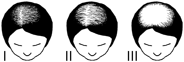 Ludwig classification stages 1-3 of female pattern hair loss. The hairline is preserved while the part at the crown progressively thins (Keministi, CC0)
Not every patient fits neatly into these typical male or female patterns, however. A Korean multicenter research team therefore developed the BASP classification, which applies the same system to both sexes to record hair loss patterns in detail (Lee Won-Soo et al., 2007). The BASP classification combines a basic type (L, M, C, or U) based on the shape of the front hairline with a specific type (F or V) based on how hair density decreases at the front and crown. An analysis of 2,213 patients who visited 13 university hospitals in Korea found that M1-2 type, in which the hairline recedes on both sides, and F1-2 type, in which the front of the crown thins, were the most common patterns among Korean men.
In an epidemiological survey of the general Korean population, the prevalence of Norwood stage 3 or higher hair loss in men was 14.1%, and the prevalence of Ludwig stage 1 or higher hair loss in women was 5.6% (Paik J H et al., 2001). Notably, the same survey found that 11.1% of Korean male hair loss patients showed a so-called female pattern, with overall thinning across the top of the head rather than hairline recession.
If Hair Suddenly Falls Out in Clumps or a Round Patch Goes Bald
Not all hair thinning is androgenetic alopecia. We use a precision scalp diagnostic device to magnify and examine hair density and scalp condition, and we classify the hair loss into seven types: male-pattern, female-pattern, alopecia areata, telogen effluvium, inflammatory, traction, and scarring.
Under magnification, if hairs that have kept their original thickness are mixed with hairs that have thinned to a vellus-like fineness within the same area, this variation in hair thickness means that follicle miniaturization is underway. This variation is therefore an important diagnostic clue for androgenetic alopecia (Liu Yingzi et al., 2025). Unlike this kind of gradual thinning, if daily shedding across the whole scalp suddenly rises to handfuls at a time within a short period after childbirth, major surgery, a severe fever, or rapid weight loss, we suspect telogen effluvium first.
If a specific area has gone bald with a relatively well-defined, coin-shaped border, this round bald patch may correspond to alopecia areata, a condition involving an autoimmune mechanism. Two other types must also be told apart: traction alopecia, which develops along the margins where hair is pulled by a habit of tying it back tightly, and scarring alopecia, in which damaged scalp tissue leaves the follicle openings visibly gone. When scalp changes such as thick flaking, redness, or itching appear together with the hair loss, we classify it as inflammatory hair loss and treat the scalp condition first.
In female patients, if hair thinning is accompanied by facial or body acne, hirsutism (coarsening of body hair), or irregular menstrual cycles, they should be evaluated for an endocrine disorder related to excess androgen in the body (PMID 30785992). If hair sheds rapidly over the course of just a few months, if a well-defined round bald patch develops, or if the scalp shows swelling, oozing, or severe pain, you should first have a medical examination to rule out other hair loss conditions, rather than starting with general scalp care.
When Are Medications, Injections, and Head Spas Used?
The medical literature treats oral finasteride for male-pattern hair loss and topical minoxidil for male- and female-pattern hair loss as the main drug treatments (Liu Yingzi et al., 2025). Among these, finasteride works by inhibiting type 2 5-alpha reductase in the body, which lowers DHT concentrations in the blood and scalp (Kaufman K D et al., 1998).
Guri Top Bone Clinic's Head Reset Fit program uses scalp autologous blood injection as its core treatment, applied to scalp areas where hair follicles are still present. We draw a small amount of the patient's own blood and centrifuge it to prepare a solution concentrated with regenerative cells and growth factors. Without any culturing step, we inject it on the day of the blood draw into the scalp around the dermal papillae, dividing it across several points at a consistent depth with a Dermashine injector. This scalp autologous blood injection is performed to support the growth activity of remaining hair follicles, and our clinic materials also describe a method that uses the same concentration process for intravenous infusion.
 Guri Top Bone Clinic's centrifuge equipment that concentrates regenerative cells and growth factors from the patient's own blood
A meta-analysis published by Anitua and colleagues pooled 43 randomized PRP studies on hair loss with 1,877 participants. In this meta-analysis, the activated PRP group showed increased hair density compared with the placebo group, but the hair thickness results did not show a statistically significant difference between the groups (Anitua Eduardo et al., 2025).
Depending on the hair loss type and scalp condition found on magnified scalp diagnosis, Guri Top Bone Clinic may also choose one of placenta, Benev, peptide, or exosome injections. During a single visit under the Head Reset Fit program, the treatment proceeds in this order: Head Manual Technique, scalp injection, Lympha-TE, and TMS. The head spa we run alongside it is also set up differently for each hair loss type: for the oily or combination scalp of male-pattern hair loss with heavy sebum production, we use a mildly acidic deep-cleansing shampoo for oily scalps, and for the dry or sensitive scalp of female-pattern hair loss, a mildly acidic, low-irritation shampoo. Before a scalp injection, we check whether the patient is taking any medication that affects blood clotting. Because the injection site may feel stiff or turn red for a few days afterward, we ask patients to pause hair dyeing, perms, and strong scalp massage on the day of the procedure.
What Happens If It's Left Untreated for Two Years?
Androgenetic alopecia does not stop on its own once it begins; it progresses gradually. In a representative long-term clinical trial reported in the literature, Kaufman and colleagues randomly assigned 1,553 men aged 18 to 41 with male pattern hair loss and tracked how their hair changed over two years. In this study, participants who took a placebo containing no active ingredient showed a continuous decline in hair count throughout the entire two-year observation period (Kaufman K D et al., 1998).
In contrast, when the group that took 1 mg of finasteride daily, a treatment described in the literature, was compared with the placebo group, hair count at the 1-inch-diameter (5.1 cm² area) measurement site on the crown was on average 107 hairs higher at 1 year and 138 hairs higher at 2 years (Kaufman K D et al., 1998). Given that the average hair count at this measurement site was 876 at the start of the study, a substantial density gap opened up within two years depending on whether treatment was received.
Hair count is maintained or increased with treatment because the hair follicles have not yet disappeared completely and can still produce thick hair again. In fact, Whiting's study analyzed scalp tissue from patients who responded to finasteride treatment and reported clinical and histological findings showing that small, miniaturized follicles enlarged and recovered again within even a single hair cycle (Whiting D A, 2001). Long-term treatment of androgenetic alopecia therefore aims to suppress progression so the hair loss does not worsen further, and to help hair grow well from the follicles remaining on the scalp so that density and thickness can recover.
If hair loss has progressed for a long time and follicle miniaturization has run to completion such that the follicles themselves have disappeared from an area, autologous hair transplantation, a treatment established in the literature, may be considered. Healthy follicles on the back of the head (occipital region) are relatively less affected by androgens and tend to keep their thickness throughout life; autologous hair transplantation is a surgical treatment that harvests these follicles and relocates them to balding areas where hair is absent (Liu Yingzi et al., 2025). However, the surrounding hair that was already present may continue to be affected by androgens and become thinner even after the transplant.
Sung-Woog Lee · Medical Director · Board-Certified Neurologist · Guri Top Bone Clinic
References
- Liu Yingzi, Tosti Antonella, Wang Etienne C E, Heilmann-Heimbach Stefanie, Aguh Crystal, Jimenez Francisco (2025). Androgenetic alopecia.. Nat Rev Dis Primers. PMID: 41068174
- Kaufman K D, Olsen E A, Whiting D, Savin R, DeVillez R, Bergfeld W (1998). Finasteride in the treatment of men with androgenetic alopecia. Finasteride Male Pattern Hair Loss Study Group.. J Am Acad Dermatol. PMID: 9777765
- Whiting D A (2001). Possible mechanisms of miniaturization during androgenetic alopecia or pattern hair loss.. J Am Acad Dermatol. PMID: 11511857
- Paik J H, Yoon J B, Sim W Y, Kim B S, Kim N I (2001). The prevalence and types of androgenetic alopecia in Korean men and women.. Br J Dermatol. PMID: 11453914
- Carmina Enrico, Azziz Ricardo, Bergfeld Wilma, Escobar-Morreale Héctor F. Female Pattern Hair Loss and Androgen Excess: A Report From the Multidisciplinary Androgen Excess and PCOS Committee. J Clin Endocrinol Metab. PMID: 30785992
- Lee Won-Soo, Ro Byung In, Hong Seung Phil, Bak Hana, Sim Woo-Young, Kim Do Won (2007). A new classification of pattern hair loss that is universal for men and women: basic and specific (BASP) classification.. J Am Acad Dermatol. PMID: 17467851
- Anitua Eduardo, Tierno Roberto, Alkhraisat Mohammad Hamdan (2025). Platelet-Rich Plasma in the Management of Alopecia: A Systematic Review and Meta-Analysis of Clinical Evidence.. Dermatol Ther (Heidelb). PMID: 40944844
Frequently Asked Questions
Q. Can male-pattern or female-pattern hair loss occur even without a family history of hair loss?
Male-pattern or female-pattern hair loss can occur even without a clear family history of hair loss. Rather than judging by family history alone, we record where the hair is thinning and how fast it is progressing, and we check for differences in hair thickness with a magnified scalp examination.
Q. When is a hormone-related evaluation needed for female-pattern hair loss?
If hair loss occurs along with irregular periods, hair becoming coarser, or acne that has suddenly worsened, a hormone-related evaluation may be needed. We check when these changes started and which medications you are taking, then decide which tests are needed.
Q. How should I take photos at home to compare before and after treatment?
Photograph the crown and the hairline under the same lighting and from the same distance, keeping the part position and shooting angle consistent each time. How wet the hair is or how long it has grown changes how much scalp shows through, so take the photos under the same conditions as much as possible.
Q. If my hair loss stage stays the same, can treatment still produce a change?
Hair loss stages are broad categories that group ranges and patterns, so hair thickness or density can still differ within the same stage. After treatment, we compare not only whether the stage has changed but also close-up photos of the same area and measured hair thickness and density.